Showing posts with label DNA. Show all posts

Wednesday 28 August 2024

New Video Posted: Introduction to Genetic Engineering: Ethics, Science, and Innovation

I have posted a video - Introduction to Genetic Engineering: Ethics, Science, and Innovation.

Genetic engineering often evokes strong emotions and heated debates. When people hear the term, they might immediately think of genetically modified organisms (GMOs), "Frankenstein foods," or the ethical dilemmas surrounding altering life at its most fundamental level. But what exactly is genetic engineering, and what are the implications of this technology?

In its simplest form, genetic engineering directly manipulates an organism's DNA to achieve desired traits. Humans have used genetic engineering through selective breeding to cultivate crops and livestock that yield better produce and more reliable outcomes for centuries. But today’s technology allows scientists to bypass traditional breeding processes and make precise changes to DNA in a laboratory setting. This raises a profound question: just because we can modify life in this way, should we?

In the video, I look at the ethical and scientific complexities of genetic engineering and wonder where society should draw the line. Is it acceptable to engineer a potato or a chicken for better production? What about a cow? How about a human? These questions are not just theoretical but have real-world implications as technology continues to advance.

I also discuss how genetic engineering can be further divided into two broad categories of genetic engineering: research and applications. Research involves using genetic engineering to understand biological systems and diseases, while applications focus on improving crops, livestock, and human health. The ethical dilemmas become particularly acute when considering human health. Should genetic modifications be limited to somatic cells (which don’t get passed on to offspring), or is it ethical to alter germ cells, thereby affecting future generations?

Finally, I wrap up the video by explaining the difference between transgenic and non-transgenic organisms. Transgenic organisms have DNA from a different species introduced into their genome, while non-transgenic organisms involve changes made to the organism’s own DNA. But this distinction leads to another intriguing question: Is DNA truly species-specific?

Additional Resources

Tuesday 27 August 2024

New Video Posted: Site-Directed Mutagenesis Explained | Understanding the Basics

In this video - Site-Directed Mutagenesis Explained | Understanding the Basics - I look at how you can mutate DNA in the lab.

Blog Bonus: Free information sheet summarising the video and defining the key terms - download.

Site-directed mutagenesis is a powerful technique in molecular biology that can introduce specific mutations into a DNA sequence. The method has been instrumental in understanding how proteins function and how genes are regulated. 

The Process of Site-Directed Mutagenesis

Site-directed mutagenesis involves several key steps:

  1. Preparation of DNA: The DNA sequence of interest is either cloned into a plasmid or selected from a plasmid library.
  2. Designing the Primer: A primer is designed to bind specifically to the region of DNA where the mutation is desired. This primer is about 20 bases long, with one crucial difference—it contains the new base that will introduce the mutation. More information on Primer Design.
  3. Creating a Single-Stranded DNA Template: The double-stranded plasmid DNA is converted into a single-stranded form. This single strand serves as the template for the primer to bind.
  4. DNA Synthesis and Ligation: Once the primer is bound, DNA polymerase synthesises the complementary strand, incorporating the new base. DNA ligase is then added to seal the DNA strand, completing the synthesis.
  5. Introduction into Bacteria: The newly mutated plasmid is introduced into bacteria, where the bacteria's natural DNA repair mechanisms take over. These mechanisms either repair the mismatched DNA back to its original form or incorporate the new mutation.

Applications of Site-Directed Mutagenesis

The ability to change a single base in a DNA sequence provides scientists with a powerful tool to explore how specific mutations affect protein function and gene expression. This technique has enabled numerous studies, allowing researchers to pinpoint individual amino acids' roles in proteins and dissect complex genetic regulatory networks.

From Site-Directed Mutagenesis to CRISPR

While site-directed mutagenesis has been a common lab technique for decades, the advent of CRISPR technology has revolutionised the field of genetic engineering. CRISPR offers a more efficient and precise method for making targeted genetic changes, but the foundational principles of mutagenesis laid the groundwork for these modern advances.

Blog Bonus: Free information sheet summarising the video and defining the key terms - download.

Additional Resources

Friday 23 August 2024

New Video Posted: DNA Sequencing: Sanger Method and Beyond Explained

In the video DNA Sequencing: Sanger Method and Beyond Explained, I explain the Sanger method and look at some of the new approaches and methods that can speed up the DNA sequencing process.

Blog Bonus: Free information sheet summarising the video and defining the key terms - download.

DNA sequencing allows scientists to decode the genetic information that dictates everything from the colour of our eyes to how our cells function. One of the most widely used laboratory methods for DNA sequencing is the Sanger method, also known as the chain-terminating dideoxynucleotide method.

The Basics of Sanger Sequencing

The Sanger sequencing process begins like a PCR (Polymerase Chain Reaction), with a template DNA that you want to sequence, a single primer (and not two primers as in PCR), DNA polymerase, and nucleotides (dNTPs). However, the method also includes special nucleotides called dideoxynucleotides (ddNTPs), which play a key role in the sequencing process.

In the sequencing method, the reaction goes through 25-30 cycles of denaturing, annealing and extension, just as you would in PCR. The DNA polymerase builds new DNA strands during the reaction and adds nucleotides to the growing chain. The twist comes with the ddNTPs; when one of these is incorporated, the chain is terminated, and no further nucleotides can be added. Each ddNTP is tagged with a different fluorescent marker corresponding to one of the four DNA bases (A, T, C, or G). This allows the sequence to be read by analysing the fluorescent tags after the reaction.

Once the reaction is completed, the next step is separation. The DNA fragments are separated using gel electrophoresis. As the fragments move through the gel, a laser scans the gel for the fluorescent tags, and the sequence of the DNA is determined by the fluorescence colour at each position.

While the Sanger method has been incredibly valuable, it has limitations. It can only read between 500 to 1000 bases per reaction, making it labour-intensive and unsuitable for large-scale projects.

Moving Beyond Sanger: Advanced Sequencing Methods

As the need for faster and more efficient sequencing has grown, new methods have been developed. One such method is shotgun sequencing, where the genome is broken into random segments. Each segment is sequenced individually using the Sanger method, and then the fragments are pieced together like a puzzle to reconstruct the full genome.

Another significant advancement was Next-Generation Sequencing (NGS), also known as massively parallel sequencing. NGS technologies have revolutionised genomics by allowing the simultaneous sequencing of many DNA molecules. This high-throughput approach can detect the sequence using either fluorescent nucleotides or pH changes.

Finally, we have Third-Generation Sequencing methods, which include techniques like nanopore sequencing. In nanopore sequencing, DNA is passed through a nanopore, and the sequence can be determined by detecting changes in electrical current.

Blog Bonus: Free information sheet summarising the video and defining the key terms - download.

Additional Resources

Wednesday 21 August 2024

New Video Posted: DNA Libraries: Genomic vs. cDNA - Key Differences and Applications

 I have posted a video on the key differences between genomic DNA (gDNA) and complementary DNA (cDNA) libraries - DNA Libraries: Genomic vs. cDNA - Key Differences and Applications.

Blog Bonus: Free information sheet summarising the video and defining the key terms - download.

DNA libraries are essential tools for researchers to study and manipulate genetic material. If you're working in a lab, you'll likely encounter two main types of DNA libraries: genomic DNA (gDNA) libraries and complementary DNA (cDNA) libraries. Though both serve important roles in research, they are fundamentally different in their composition, creation, and applications. 

What Are DNA Libraries?

A DNA library is a collection of DNA sequences cloned into vectors, small pieces of DNA that can carry foreign DNA into a host cell, such as bacteria. These libraries allow scientists to store, access, and manipulate specific DNA sequences for various research purposes.

Genomic DNA (gDNA) Libraries

A genomic DNA library is created from the complete genomic DNA of an organism. This means it contains all the genetic material necessary to build that organism. In eukaryotes, this includes both the coding regions (exons) and the non-coding regions (introns) of genes. 

How is a gDNA Library Made?

  1. DNA Extraction: Genomic DNA is extracted from cells.
  2. DNA Fragmentation: The extracted DNA is cut into smaller fragments using restriction enzymes.
  3. Cloning: These fragments are then cloned into plasmids, which are circular DNA molecules.
  4. Transformation: The plasmids are inserted into bacteria, which replicate the DNA fragments, creating a library.

When to Use a gDNA Library?

gDNA libraries are ideal when studying the full structure of genes, including regulatory elements, or when exploring gene functions across the genome.

However, there are some limitations. Because a gDNA library contains both introns and exons, it can complicate the task of isolating and studying the sequences that actually code for proteins. Additionally, genes in a gDNA library may be fragmented across multiple clones, making it challenging to reconstruct the complete gene sequence through sequence alignment.

Complementary DNA (cDNA) Libraries

cDNA libraries are derived from messenger RNA (mRNA) in a cell. This means that a cDNA library only contains the sequences actively expressed as proteins when the library is made. Therefore, the content of a cDNA library depends on the type of cell, the time of day, and the cell's conditions.

How is a cDNA Library Made?

  1. mRNA Isolation: Cells are lysed, and the mRNA is purified from the lysate using affinity chromatography, which often involves oligo-dT beads that bind to the poly-A tails of mRNA molecules.
  2. Reverse Transcription: Using reverse transcriptase, the mRNA is used as a template to synthesise complementary DNA (cDNA). This gives a mRNA/DNA molecule.
  3. RNA Removal: The original mRNA is removed from the mRNA/DNA molecule using an enzyme (RNase H). The now single-stranded cDNA is converted into a double-stranded DNA molecule using DNA polymerase.
  4. Cloning: The cDNA is cloned into plasmids and inserted into bacteria.
Isolating mRNA can be tricky because it is easily degraded by enzymes released during cell lysis or by RNases in the environment. Moreover, because cDNA libraries only reflect the genes being expressed at a specific time, they may not provide a complete picture of an organism's genome.

When to Use a cDNA Library?

cDNA libraries are particularly useful for studying gene expression, identifying specific mRNA sequences, and producing recombinant proteins.

Choosing the Right Library

It is essential to understand the distinctions between gDNA and cDNA libraries. A gDNA library is the right choice if the goal is to study the entire genome or understand gene regulation. However, a cDNA library must be used if you're interested in the proteins a cell produces or need to work with specific mRNA sequences.

Blog Bonus: Free information sheet summarising the video and defining the key terms - download.

Additional Reading

The video was produced with help from the following resources:

Monday 19 August 2024

New Video Posted: PCR Primer Design: Tips for Accurate DNA Amplification

I have posted a video on PCR primer design - PCR Primer Design: Tips for Accurate DNA Amplification.

Blog Bonus: Free information sheet summarising the video and defining the key terms - download.

Polymerase Chain Reaction (PCR) is an important lab technique that allows scientists to amplify specific DNA sequences. However, the success of a PCR largely depends on correctly designing the PCR primers. 

Why Primer Design Matters

The importance of primer design in PCR cannot be overstated. If your primers are poorly designed, the DNA amplified during the reaction might not be the correct region of DNA, or you will get a low product yield. To avoid such issues, you must carefully consider three primary factors when designing primers: 

  1. Melting temperature (Tm)
  2. Disruptive secondary structures
  3. Specificity of the primers

1. Melting and Annealing Temperatures

Two crucial temperatures play a role in PCR primer design:

  1. the melting temperature (Tm)
  2. the annealing temperature
The Tm is the temperature at which 50% of the primer would dissociate from a double-stranded to a single-stranded form. The primers bind to the denatured DNA template during each PCR cycle at the annealing temperature. Typically, the ideal Tm is 55-65 °C, and the annealing temperature is set 3-5 °C below the Tm to ensure optimal primer binding.

In the lab, you would use a computer program to determine the Tm of a primer. The program usually uses the nearest neighbour method to calculate the Tm accurately. This method considers the specific sequence of bases and the concentrations of components in the reaction.

2. Disruptive secondary structures

It is essential to ensure your primers won’t form problematic secondary structures like hairpin loops or primer-dimer pairs. Hairpin loops occur when a primer folds back on itself, creating a loop that can be difficult to melt during the PCR process, leading to inefficient amplification. Primer-dimer pairs form when two primers bind to each other instead of the target DNA. This can result in an inefficient reaction and the production of the wrong DNA.

In the lab, you would use a computer program to test for hairpin loop formation and primer-dimer pairs.

3. Specificity of the primers

A standard PCR primer is usually 20 to 30 bases long and has an ideal Tm of around 55-65 °C. The Tm and the length of the primer help ensure specificity. 

Finally, it is advisable to perform a BLAST search with your primer sequences, which can help confirm that they do not have unintended matches with other sequences in the species. If the DNA species is not human, a BLAST search against a human database should be performed to check for the possibility of a result if the reaction is contaminated with human DNA.

Advanced Techniques: Overcoming Challenges in PCR

In some cases, even with well-designed primers, you might need help getting the correct DNA to amplify, especially when dealing with low amounts of template DNA or needing to add restriction sites to your product. To address this, you can use a nested primer approach. This involves using two sets of primers, one set falling inside the other, to improve specificity and yield through a two-round PCR process.

For adding restriction sites, primers are designed with the desired restriction site sequences at the 5’ end. However, this introduces a mismatch between the primer and the template DNA. To compensate, the initial PCR cycles are performed at a lower annealing temperature to help the primers bind more effectively. Once the restriction site is incorporated into the amplified DNA, the annealing temperature is raised to the standard level for subsequent cycles. A GC clamp at the 3’ end of the primer, where the last two bases are guanine (G) or cytosine (C), can also help improve binding due to the stronger hydrogen bonds these bases form.

Blog Bonus: Free information sheet summarising the video and defining the key terms - download.

Additional Reading

The video was produced with help from the following resources:

Friday 3 May 2024

DNA - that's a wrap - 14 DNA revision videos

OK, so that is a wrap on DNA, a topic of immense significance in the world of biology and genetics.

Over the past few weeks, I have released fourteen revision videos on DNA, with accompanying posts on here and with information sheets accompanying each video (you can find a link to the information sheets in the links below).

In the videos, I have covered:

I then moved on to the all-important three Rs of DNA - replication (copy), repair and recombination:
Next, I asked the important question of how cells regulate the processes of transcription and translation:
I finished off the series by looking at some lab techniques we use to work with DNA:

If you would like to support my blogging efforts, then please feel free to buy me a coffee at https://www.buymeacoffee.com/drnickm


Additional Reading

The video was produced with help from the following resources:

A Comprehensive Guide on How to Calculate the Size of a DNA Band on a Gel

How do scientists determine the size of DNA bands on an agarose gel? In this guide, I will walk you through the step-by-step process of calculating the size in base pairs of a DNA band on an agarose gel. 

Blog Bonus: Free information sheet summarising the video and the steps - download.


Introduction

When working in a lab and running an agarose gel, you may need to determine the size of the DNA fragment, and this information may be crucial for various biological research applications. 

This approach is also described in the following video:


Setting Up the Experiment

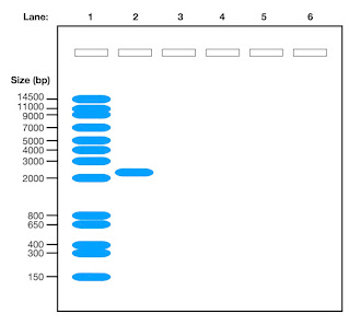
Imagine you have loaded a DNA ladder with known sizes in one lane and your DNA sample with an unknown size in another lane of the gel and you get a result that looks like this when the gel has been run.

DNA gel showing a DNA ladder and a band

Before you can calculate the size of your DNA band, you must first label the gel and collect data to create a calibration curve.

Data Collection and Analysis

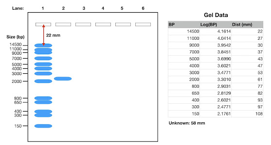
By measuring the distances the DNA bands in the ladder (see below) have moved and plotting the log values of their sizes against the distances travelled in millimetres (or you can do it in pixels), you can create a calibration curve. This curve will help you accurately determine the size of the DNA band in your unknown sample.

The image below shows the gel and the data table for the plot.

Agarose gel showing the DNA ladder and the band of unknown size, plus a table of data constructed from the gel for the grapg

From the table, you plot the calibration curve.

Calibration curve for determining the size of a band on a DNA gel

Calculating the Size of the DNA Band

After plotting the calibration curve (above) and identifying the distance your unknown band has travelled, you can use the curve to determine the size of the DNA band in base pairs. By following a simple formula involving logarithms, you can convert the log value to the actual size in base pairs.

Conclusion

Calculating the size of a DNA band on an agarose gel requires careful data collection, analysis, and interpretation. By following the steps outlined in this guide, you can confidently determine the size of DNA fragments in your samples. 

If you would like to support my blogging efforts, then please feel free to buy me a coffee at https://www.buymeacoffee.com/drnickm

Blog Bonus: Free information sheet summarising the video and the steps - download.

Additional Resources

Wednesday 1 May 2024

New video posted: DNA Cloning - how to overcome some common problems

In this video, I examine the step-by-step process of cloning DNA into plasmids and address common challenges faced in the lab. I start by preparing the DNA and then move on to using restriction enzymes like EcoRI and HindIII. I explain the importance of choosing the correct enzyme pairs to prevent self-ligation and ensure the correct orientation of the insert. I also cover the blue-white selection method to verify successful cloning and discuss using different vectors for larger DNA segments. 


If you would like to support my blogging efforts, then please feel free to buy me a coffee at https://www.buymeacoffee.com/drnickm

Blog Bonus: Free information sheet summarising the video and defining the key terms - download.
 

Additional Reading

The video was produced with help from the following resources:

New video posted: DNA Cloning - how do we clone DNA in the lab?

In this video, I give a brief introduction to the subject of DNA cloning.

DNA cloning is an important lab skill that all life and biomedical science students should possess. In the video, I provide a summary of in vivo and in vitro cloning, along with the key steps, tools and methods you would use.

If you would like to support my blogging efforts, then please feel free to buy me a coffee at https://www.buymeacoffee.com/drnickm

Blog Bonus: Free information sheet summarising the video and defining the key terms - download.
 

Additional Reading

The video was produced with help from the following resources:

Monday 29 April 2024

New video posted: Translation - making proteins from DNA - decoding mRNA to make the protein

This is the second of two videos on how cells make proteins using DNA. In the first video, I looked at the first step, which is making the messenger RNA (mRNA) a process called transcription - Transcription - making proteins from DNA - the mRNA.

In this video, I will guide you through the process of producing the protein from the mRNA, also known as translation. I will look at the coding problem (how many mRNA bases do you need to code from an amino acid), the number of reading frames in a DNA molecule, and how the cell produces protein from the mRNA.


If you would like to support my blogging efforts, then please feel free to buy me a coffee at https://www.buymeacoffee.com/drnickm

Blog Bonus: Free information sheet summarising the video and defining the key terms - download.
 

Additional Reading

The video was produced with help from the following resources:

Friday 26 April 2024

New video posted: Transcription - making proteins from DNA - the mRNA

This is the first of two videos on how cells make proteins using DNA. The second video looks at how we go from messenger RNA (mRNA) to protein - Translation - making proteins from DNA - decoding mRNA to make the protein

In this video, I will guide you through the first steps in the process of producing RNA from DNA, also known as transcription. In the next video, we will take the next step and examine how we produce the protein from mRNA.

The video looks at the five steps of mRNA production:

  • initiation — activators bind upstream, often thousands of bases upstream, of the gene. The activators assemble the required proteins (the mediator, chromatin remodelling complex, the RNA polymerase and transcription factors) at the TATA box, which is a DNA sequence close to the gene
  • production — DNA is transcribed into the pre-messenger RNA
  • five prime capping — the five prime end of the pre-messenger RNA is capped with some modified nucleotides
  • splicing — introns are spliced out of the pre-messenger RNA to leave just the exons (exons provide the sequence for the protein)
  • three prime polyadenylation — addition of a polyadenylated tail to the three prime end of the pre-messenger RNA to give the final mature messenger RNA molecule

The video not only looks at mRNA production but also introduces the idea of non-coding RNA (ncRNA), which are RNA molecules that do not encode proteins. ncRNAs are essential in regulating gene expression and various cellular processes. For example:

  • transfer RNA (tRNA) — involved in protein synthesis
  • ribosomal RNA (rRNA) — involved in protein synthesis
  • microRNA (miRNA) — control gene expression
  • small interfering RNA (siRNA) — control gene expression 
  • long non-coding RNA (lncRNA) — diverse functions
  • circular RNA (circRNA) — gene regulation
  • Piwi-interacting RNA (piRNA) — genome protection
  • enhancer RNAs (eRNAs) — modulate gene activity
Finally, I cover how to write out DNA sequences, the means of the terms sense and antisense strands, and what we mean by upstream and downstream when talking about DNA and RNA molecules.

If you would like to support my blogging efforts, then please feel free to buy me a coffee at https://www.buymeacoffee.com/drnickm

Blog Bonus: Free information sheet summarising the video and defining the key terms - download.
 

Additional Reading

The video was produced with help from the following resources:

Thursday 18 April 2024

New video posted: How can DNA become damaged in the cell?

In this video, I look at how DNA can become damaged in cells and introduce four ways damage can occur:

  • deletions
  • insertions
  • substitutions
  • transitions
  • and transversions. 
I also look at exogenous and endogenous damage sources of the damage.

If you would like to support my blogging efforts, then please feel free to buy me a coffee at https://www.buymeacoffee.com/drnickm

Blog Bonus: Free information sheet summarising the video and defining the key terms - download.
 

Additional Reading

The video was produced with help from the following resources:

Wednesday 17 April 2024

New video posted: DNA replication: How does a cell make a copy of its DNA?

Have you ever been curious about the precision with which DNA is replicated in our cells, or why human pregnancies don't extend to an unimaginable sixteen years?

In my latest video, I examine the process of DNA replication. First, I explore how the DNA double helix is unwound by DNA helicase and how the unwound DNA is stabilised by Single Stranded Binding Proteins. I look at the replication fork and how DNA polymerases copy the DNA strands.

I also cover the formation of Okazaki fragments—short sequences of DNA synthesised on the lagging strand—and how DNA ligase is responsible for stitching these fragments together. Additionally, the video highlights the importance of topoisomerase in preventing the DNA from becoming overly twisted and tangled during replication.

If you would like to support my blogging efforts, then please feel free to buy me a coffee at https://www.buymeacoffee.com/drnickm

Blog Bonus: Free information sheet summarising the video and defining the key terms - download.
 

Additional Reading

The video was produced with help from the following resources:

Monday 15 April 2024

New video posted: DNA Packaging - how do we package DNA into the nucleus?

In the video, I examine how the long DNA molecule is packaged into the relatively small nucleus of a cell. I examine how DNA is broken up into chromosomes, how the DNA wraps around the histones to form nucleosomes and chromatosomes, and finally, chromatin.

Please use the form below to download a fact sheet for the video. The fact sheet contains twenty-four key facts from the video, a summary of the packing of DNA into the nucleus and the definitions of twenty-five key terms used in the video.

If you would like to support my blogging efforts, then please feel free to buy me a coffee at https://www.buymeacoffee.com/drnickm

Blog Bonus: Free information sheet summarising the video and defining the key terms - download.
 

Additional Reading

The video was produced with help from the following resources:

Thursday 11 April 2024

New video posted: How long is the DNA in a human cell?

In this video, I calculate the length of the DNA in one human cell, and my answer may surprise you.

How long do you think? 0.2 mm, less? 2 mm, 2 cm, 2 m, 2 km? Watch the video to find out.

The video walks you through the math I used to calculate the length of DNA, and then I will reveal the length it would be if the cell were the size of a football.

If you would like to support my blogging efforts, then please feel free to buy me a coffee at https://www.buymeacoffee.com/drnickm

Blog Bonus: Free information sheet summarising the video and defining the key terms - download.
 

Additional Reading

The video was produced with help from the following resources:

Monday 8 April 2024

New video posted: DNA and Genes

How many genes does a human have? Do we have more genes than a plant? Do larger organisms, such as trees, have more genes than humans?

In the video, we will look at the genetic material — DNA (deoxyribonucleic acid) — in cells and explore how many genes humans have and whether we, as complex organisms, have more genes than a tree, a potato or a tomato. What do you think?

If you would like to support my blogging efforts, then please feel free to buy me a coffee at https://www.buymeacoffee.com/drnickm

Blog Bonus: Free information sheet summarising the video and defining the key terms - download.
 

Additional Reading

The video was produced with help from the following resources:

Monday 5 October 2009

Protein sequence to DNA - Degeneracy calculation

I have had some questions about the reverse translation of protein to DNA and degeneracy...

The protein sequence is THERIGHTREADINGFRAME

It is 20 amino acids, and therefore, you will need 60 bases to encode it. So....

Protein Seq:  T  H  E  R  I  G  H  T  R  E  A  D  I  N  G  F  R  A  M  E 
DNA Seq:     ACNCAYGARMGNATHGGNCAYACNMGNGARGCNGAYATHAAYGGNTTYMGNGCNATGGAR    
Full DNA:    ACACACGAACGAATAGGACACACACGAGAAGCAGACATAAACGGATTCCGAGCAATGGAA
               T  T  G  T  C  T  T  T  T  G  T  T  C  T  T  T  T  T     G
               G        G  T  C     G  G     C     T     C     G  C
               C        C     G     C  C     G           G     C  G
                      AGG            AGG                     AGG
                        A              A                       A
Number codons: 4  2  2  6  3  4  2  4  6  2  4  2  3  2  4  2  6  4  1  2
So, 4 x 2 x 2 x 6 x 3 x 4 x 2 x 4 x 6 x 2 x 4 x 2 x 3 x 2 x 4 x 2 x 6 x 4 x 1 x 2 = 2,038,431,744 or 2 x 10⁹ possible DNA sequences would encode the protein sequence.

This is a big number; however, compared to the total number of possible DNA sequences you could have for a 60-base sequence, it is small.

The total number of DNA sequences you could have for a 60 base sequence is 4 x 4 x 4.... sixty times, or 460, which is equal to 1.3 x 1036 possible sequences. Of those 1.3 x 1036 sequences only 2,038,431,744 would encode THERIGHTREADINGFRAME. Or in percentage terms, (2,038,431,744 / 1.3 x 1036) x 100 = 0.0000000000000000000000002% (2 x 10-25%) of all the possible sequences would encode THERIGHTREADINGFRAME.

You may find the following video useful where I explain the above:


If you would like to support my blogging efforts, then please feel free to buy me a coffee at https://www.buymeacoffee.com/drnickm

Additional Resources